AFFERENT MOTOR APHASIAS
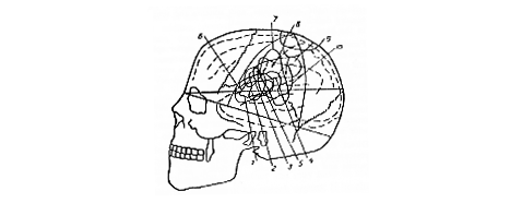
Luria distinguished two forms of afferent motor aphasia as arising from lesions of different parts of the left parietal lobe, the combined primary and secondary sensory areas vs. the tertiary sensory area.
He regarded the primary sensory area to be the pharyngeal and laryngeal portions of the postcentral gyrus; more specifically as the cytoarchitecturally defined rostral postcentral area 3, intermediate postcentral area 1 and caudal postcentral area 2 of Brodmann.
The secondary sensory area was identified in general terms as the "posterosuperior parietal area", and more specifically as the cytoarchitecturally defined preparietal area 5 and part of the superior parietal area 7 of Brodmann.
The tertiary sensory area was identified generally as the "posteroinferior parietal area" and more specifically identified with the remainder of the superior parietal area 7 and the anterior part of the supramarginal area 40 of Brodmann.
 |
|
|
|
|
Psychophysiology of the Afferent Motor Aphasias
The importance of feedback for the smooth execution of skilled movements was well accepted. For instance, it had long been recognized that the ataxic gait of a patient with tabes dorsalis resulted from a loss of kinesthetic feedback. The major subcortical input to the parietal lobe comes from the kinesthetic and somatosensory tracts. Thus its primary function is assumed to be sensory in nature. The fact that stimulation of the postcentral gyrus gives rise to movements similar to those produced by stimulation of the precentral gyrus (Penfield and Rasmussen,1950) suggested a close relationship to the motor analyzer.
The common denominator of the afferent motor aphasias was a "positional apraxia" or "ataxia of the speech apparatus", i.e., of the lingual, pharyngeal, and laryngeal musculature. A patient with a lesion of the parietal area lost the "articulatory schemata", of phonemes and words. Thus he had difficulty in finding the articulatory positions appropriate for pronouncing different phonemes. Instead of confusing phonemes that sound alike, he tended to confuse phonemes, or "articulemes", that are similar in articulation, e.g., "1", "n", "t", "d", and "s". Destruction of the kinesthetic-somatosensory cortex was thought to disrupt the selective inhibition of motor impulses arising in the primary motor cortex, which resulted in a mischannelling of impulses to the vocal apparatus. Just as visual feedback had to substitute for kinesthetic feedback in the tabetic patient, a patient with a parietal lobe lesion had to substitute auditory control for the kinesthetic control of articulation. He could not pronounce words accurately unless he could hear what he was saying.
Expressive Speech Disorders in Afferent Motor Aphasias
The most prominent defects in afferent motor aphasia involved expressive speech. The patient glided from one articulatory position to another until he found the sound he wanted. In some cases the articulatory positions remained intact, so there was no slurring of speech, but the patient had difficulty producing the appropriate configuration of the speech apparatus to produce the desired sound. He made frequent paraphasic errors, substituting similar articulatory patterns for one another. The following tests were used to evaluate the degree of control that the patient could exert over oral movements.
1. coordinate tongue and lip positions in response to verbal instructions, e.g., "Touch your upper lip with your tongue;"
2. imitate tongue and lip positions demonstrated by the examiner;
3. produce "practical actions" involving the mouth, e.g., blow out a match or spit;
4. produce symbolic movements of the oral apparatus, e.g., demonstrate lip movements appropriate to whistling;
5. speak spontaneously while the examiner listens for literal paraphasias;
6. read and imitate spoken phonemes, syllables and words.
Receptive Speech is usually intact in patients with afferent motor aphasia.
Reading and Writing are affected in patients who, prior to injury, were only able to read and write by articulating words to themselves as they did so.
The predominant signs of afferent motor aphasia depend to some extent on the site of the lesion. About 40% of cases with lesions in the primary and secondary sensory areas involve difficulty in controlling movements of the articulatory apparatus that is so gross as to border on dysarthria. Neither practical, symbolic, nor articulatory movements are performed with skill. These difficulties may be accompanied by "afferent paresis" or loss of fine movements in the right hand.
In 95% of cases with lesions of the tertiary sensory area dysphasia is apparent. There may be no slurring, but literal paraphasias are common. Imitative and practical actions of the oral musculature may be intact, but symbolic movements are impaired. Astereognosis, ideomotor apraxia ideational apraxia frequently accompany these signs.