EFFERENT MOTOR APHASIAS
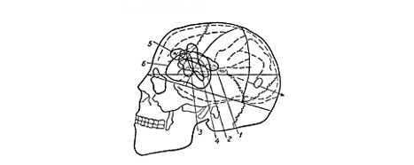
Efferent motor aphasias result from damage to a portion of the frontal lobe that includes part or all of six different cortical areas as defined by Brodmann on the basis of cytoarchitecture, namely, areas 6 (more specifically areas 6a-alpha and 6a-beta of the Vogts), 8 (more specifically 8a-beta of the Vogts), 44, 45, 46, and 47. Luria divided them into three groups that he identified with different syndromes of efferent motor aphasia.
He identified the superior premotor area or secondary motor area with the agranular frontal area 6 and the intermediate frontal area 8 of Brodmann.
The inferior premotor area he identified with the opercular area 44 of Brodmann, Broca's area.
He used the term frontal area to refer to the triangular area 45, middle frontal area 46, and orbital area 47 of Brodmann.
 |
|
|
|
|
Psychophysiology of the Efferent Motor Aphasias
The common denominator of aphasias arising from frontal lobe lesions is impairment of the seqential pattern of speech, or loss of the "sequential dynamic schemata" of speech. Luria attributed two types of symptom to the loss of sequential schemata, viz, (1) loss of the smoothness with which speech is produced and (2) perseveration. Perseveration is thought to result from a "denervation difficulty", i.e., once a phoneme or word has been uttered it must be denervated, or inhibited, before the next element can be uttered. The signs of efferent motor aphasia vary depending on the site of cortical damage. Likewise, the tests used to elicit signs of dysfunction depend upon the site.
Superior Premotor Area: Tests and Signs of Dysfunction
Luria concluded that the function of the superior premotor area is to integrate impulses arising in different parts of the primary motor area (gigantopyramidal area 4 of Brodmann) to produce smooth movement patterns. This area is necessary for the development of skilled movements ... "automatic movement sequences" or "kinetic melodies."
The most prominent sign of damage to this area is loss of the automaticity of speech. Spontaneous speech is interrupted by word-searching which need not involve primarily nouns; the patient can name objects. The grammatical complexity of speech is reduced. Tests for damage to the superior premotor area are not restricted to verbal tasks; they include the following.
1. Reciprocal Coordination. The hands are placed on the table, one with palm down, the other in a fist; the patient is instructed to reverse their positions repeatedly and smoothly. Another technique is to have the hand positioned as though playing a piano and to tap the table with the fingers in a certain repetitive sequence, e.g., 1-2-1-5-1-2-1-5,...etc.
2. Imitation of Rhythm. The examiner taps a rhythm, e.g., // // //, and the patient attempts to imitate it. With a premotor lesion he may fail to do so. He may reproduce the first pattern correctly but then fail to reproduce a second pattern, such as /// /// /// because of perseveration of the first response. (Care must be taken to distinguish failure resulting from motor impairment from failure due to an auditory defect). If the defect is premotor in origin, the patient often recognizes his error but is unable to correct it.
3. Imitation of Verbal Sequences. Having repeated the sequence "house-bird-cat" the patient may be unable to shift to the sequence "bird-house-cat" -- an example of verbal perseveration.
Inferior Premotor Area: Tests and Signs of Dysfunction
Luria regarded the function of the inferior premotor area as similar to that of the superior area, except that it is more specifically concerned with speech activity.
The predominant signs of damage to "Broca's area" are also loss of the "automaticity of speech" with the emergence of perseveration. There may be a loss of "dynamic schemata" at the phonemic, word, or sentence level. If disruption occurs at the phonemic level the patient may be totally aphasic. If it is less severe, some speech is retained, but the patient loses his "feel for the language," i.e., his speech lacks normal intonation and is composed of sentence fragments rather than connected phrases. The "predicative aspect" of speech suffers more than the "nominative aspect", i.e., verbs and adjectival, adverbial, and prepositional phrases are affected more than nouns. This gives rise to the so-called "telegraph style" of speech, e.g., "I came... Moscow...hospital ... doctor ... questions..."
Frontal Area: Tests and Signs of Dysfunction
He regarded the frontal area as particularly necessary for the formulation of the intention to say things.
Occasionally a patients with frontal lesions were found to be aphasic. They failed to speak, spontaneously; seldom using speech as a means of communication. If some speech was retained they might complain that they were unable to organize their thoughts or that they forgot what they intended to say before the sentence was half out.
The investigation of inferior premotor and frontal area lesions primarily involves tasks that require the patient to reproduce a story, describe a previous experience, make up a story on a given theme, or put together a story using ready-made sentences provided by the examiner in written form. He found that a patient unable to recount a story on his own was sometimes able to do so if the examiner merely supplied connectives and transitional phrases at the appropriate times, e.g., "Once upon a time", "and then", "after that", etc.