SEMANTIC APHASIAS
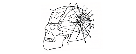
Semantic aphasias usually result from damage the region of the left hemisphere where the occipital lobe, the temporal lobe and the parietal lobe meet. Although semantic aphasia cannot be considered primarily a visual defect, it possesses a visual component. Thus, for the sake of completeness, Luria's conception of the cortical organization of the visual system is presented here. Six cytoarchitecturally defined areas are involved: areas 17, 18, 19, 37, 39 and 40 of Brodmann.
The primary visual area is the striate area 17 defined by Brodmann on the basis of cytoarchitecture.
The secondary visual area includes the parastriate area 18 and part of the peristriate area 19 of Brodmann.
The temporo-parieto-occipital area was regarded by Luria as including the angular area 39 and parts of the supramarginal area 40, the peristriate area 19, and occipitotemporal area 37 of Brodmann; Luria also referred to it as the "TPO-tertiary zone".
 |
|
|
|
|
Psychophysiology of the Semantic Aphasias
The left primary visual area receives fibers in somatotopic distribution from the left half of each retina. Thus, it receives the input necessary for a point-to-point representation of the right half of the visual field. Penfield and others had shown that stimulation in that area produced a flash, or "phosphene". Resection of the area was known to produce a left homonymous hemianopsia, i.e., patient does not respond to visual stimuli in the right half of the visual field. The limits of such a visual field defect are determined by the "confrontation" method or by perimetry.
The secondary visual area is involved in the perception of visual patterns, viz, in the recognition of so-called "simultaneous patterns", as opposed to the "sequential patterns" with which the premotor area is concerned. Stimulation of this area may produce formed images; ablation leads to abnormal control of eye movements and instability of vision, so that when patients read they may jump lines or begin reading in the middle of the page. Spatial perception is impaired, and the field of visual attention may be constricted, i.e., the patient may be able to see only one or two objects at a time, regardless of their size, background, or position in the visual field (simultagnosia). The following tests were used to demonstrate damage of the secondary visual area.
1. reading: the patient may skip from line to line or read haltingly as he perceives only one word at a time.
2. tests for constriction of the field of visual attention: the patient is presented with a card bearing a circle and a square and is instructed to place a dot between the two figures; if he is unable to see the two simultaneously, he cannot place a dot between them.
3. tests of the ability to (a) imitate hand positions demonstrated by the examiner, (b) draw a floor plan of the ward or (c) draw a rough map of a familiar area, may be used to demonstrate impairment of spatial perception.
4. name objects the outlines of which have been crossed out may demonstrate an inability to recognize figure ground relationships.
The temporo-parieto-occipital area is phylogenetically one of the newest areas of the human cortex and constitutes the "zone of overlap" between. the cortical portions of the auditory, somesthetic-kinesthetic, and visual analyzers. Penfield had reported that electrical stimulation of this area occasionally produced deja vu, a visuo-auditory hallucination or the "reliving" of an old experience. Destruction of the area produced a number of signs that Luria judged could be traced to a loss of "simultaneous schemata", i.e., to loss of the memory for, or ability to recognize, patterns in a group of auditory, visual, and/or somesthetic stimuli occurring simultaneously. This concept was similar to that of other investigators. According to Goldstein the antero-occipital area played an important role in the "transformation of sequential recognition processes into instantaneous recognition." According to Head the area was necessary for the organization of individual "traces" into over-all, "simultaneous systems." Luria found that the most prominent signs of damage to this area were spatial disorientation, loss of the "predicative" aspect of speech, agraphia, and alexia.
1. Spatial disorientation was demonstrable by a number of tests.
- a. Shown the north and south poles of a compass, the patient
was unable to tell which pole represented East and which West.
- b. In drawing a map he positioned landmarks according to associations that occurred to him impulsively rather than according to conventional geographic coordinates.
- c. He might be unable to tell which was his left hand and which was his right.
- d. He was unable to reproduce hand positions demonstrated by the examiner sitting opposite him, presumably because it required mentally reversing the positions as he saw them.
- e. He was unable to reproduce the orientation of simple match-stick patterns laid out by the examiner opposite him for the same reason.
- f. He might be able to copy geometrical figures in the same orientation as they appeared on a paper before him, but if they were shown and taken away, he drew them in random orientation.
- g. In attempting to write he might construct individual letters upside down or backwards.
2. The speech disturbances of semantic aphasia were said to arise from the facts that (1) the patient was unable to deal with grammatical constructions and sentences as a whole and (2) he lost the meanings of words that derive from their relationship to general frames of reference, i.e., he retained the denotative or "nominative" meanings of words but lost the connotative or "predicative" meanings.
Loss of the ability to handle grammatical constructions and to deal with sentences as a whole was shown by the following tests:
- a. The patient might comprehend long sentences in which the
grammar was simple but fail to comprehend shorter sentences which
involved subordinate clauses or prepositional phrases.
- b. Instructed to "draw a circle under a square", he might draw a circle and, under it, draw a square. The sequence of action in this case matched the order of words in the instruction rather than its meaning, which was conveyed by the grammatical structure. This phenomenon was referred to as "receptive agrammatism."
- c. He might be able to name the days of the week in correct order, but be unable to recite them backwards or answer such questions as, "What day comes before Wednesday?" Luria regarded this as evidence that he could not conceptualize or operate on the series as a unit to derive information in the form required by the task.
3. Loss of the predicative aspect of words led to:
- a. inability to comprehend metaphor
- b. nominal aphasia. The form of nominal aphasia seen with temporo-parieto-occipital lesions differed from that produced by damage to the postero-inferior temporal lobe in that prompting helped. Often the examiner needed only provide the first letter of the word sought, and the patient recalled it. Luria concluded that in this case the patient had not lost the auditory schema of the word but the system of associations that, for the normal person, mediate the process of recall.