This is a publication in Web format of brainstem sections, some of which were originally published in Smith-72 in the Journal of Comparative Neurology (Wistar Institute Press, now Wiley-Liss, Inc.)
The Web version of the atlas consists of 72 digitized images of coronal sections of the rhesus brainstem in stereotaxic coordinates relative to the orbitomeatal plane. The zero of all three dimensions is at earbar 0 (eb 0), the midpoint of the interaural line. This version of the atlas includes new images of the previously published 19 coronal sections stained by Cresyl violet for Nissl substance as well as 53 intervening sections at 100 to 200 mu intervals.
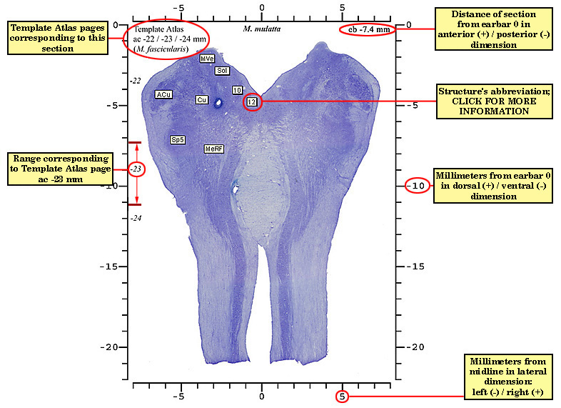
The printout of a section as it first appears on your screen bears an image magnified approximately 8:1 relative to brain size in the living subject. To print pages for an atlas that is satisfactory for stereotaxic purposes, simply click your browser's PRINT button for each section to be included. You may want to include only the sections that appeared in the original publication; they are indicated by bold type in the sidebar and are labeled to indicate the location of major landmark structures. There are 19 labeled sections occurring at 0.3 to 0.8mm intervals.
You can obtain images showing greater structural detail by clicking the Zoom 2.5x and Zoom 5x buttons in the sidebar. Zoom 2.5x provides a 20:1 magnification and will be suitable for most purposes. The precise magnification of a given printout can be determined by measuring the distance in millimeters between ticks on the scale, which corresponds to 1mm in the brain of the intact subject. The image of a full Zoom 2.5x section occupies about 60 cm x 90 cm (~2 ft x 3 ft). The most efficient means of printing a region of interest from an image of that size is to download the image file into an image processing program such as Adobe Photoshop, where it can be manipulated and cropped as desired. If your operating system is Microsoft Windows, right click on the image and select "Save picture as…"
Zoom 5x provides the maximal resolution obtainable without distortion by pixelation (40:1). It produces an image about 120 x 180cm (4 x 6 ft). In our experience, this magnification does not reveal much greater structural detail than Zoom 2.5x, but it may be useful for some purposes. Users with a computer of limited resources or a modem that transmits at less than 256 baud may find downloading and scrolling the 5X images to be very slow.
See the publication Smith-72 for a full description of the specimens and methodology used to prepare and register the sections to the stereotaxic space.
Many of the sections, which were cut and mounted on slides in the early 1970's, were faded and distorted by crystallization of the mounting substance. To produce the web atlas, the coverslip of each section was removed, the tissue was rehydrated and restained with Cresyl violet, and a new coverslip was applied. Photomicroscopy was performed using a digital camera (Sony DKC5000 3CCD) coupled to a microscope (Nikon Eclipse E800) and a motorized stage controller (ProScan H101).
Images were acquired using Universal Imaging Corporation MetaMorph and software developed by the Bioengineering Division of the Washington Regional Primate Research Center to allow semiautomatic tiling of multiple microscopic fields into a single image. The image of each section was digitized at 72 pixels/inch. The images with scales, labels, borders, etc. were generated using Adobe Photoshop on a Gateway 2000 PC running Microsoft Windows 98.
Labels for the major landmark structures have been applied to the same structures of the same sections as in the original printed atlas. Labeled sections are indicated by the bold font of section names in the sidebar, e.g., eb -9.7 for the section 9.7mm posterior to the earbars . For interoperability with other parts of BrainInfo, the labels have been changed from the Latin of the original publication to English. Click the label abbreviation for the full name of the structure and more information about it. If a label obscures a structure one can view the structure by clicking Zoom 2.5x and scrolling to the structure or by clicking anterior and posterior to view the structure in adjacent sections.
The scale was generated by measuring the distance on all sections between registration landmarks, which had been created by the passage of an electrode at 10mm intervals in the vertical dimension, and dividing by 10mm to obtain a scale factor in units/mm for each section. We calculated the mean scale factor across sections and used it to create identical scales for all of the images. The magnification of the images on your computer screen depends on the display setting of your computer. You can determine it by measuring the distances in millimeters between ticks on the scale; the distance between ticks corresponds to 1mm in the brain of the intact animal.
Contributor |
Affiliation* |
Contribution |
| John R. Bolles | Health Sciences Center for Educational Resources | Web atlas developer (2001) |
| Douglas M. Bowden | Washington Regional Primate Research Center and Department of Psychiatry and Behavioral Sciences | Designed and edited Web version of the atlas (2000-2001) |
| Andra Erickson | Washington Regional Primate Research Center | Histologist for rehydration and restaining of slides (2000) |
| Kenneth G. Kastella | Washington Regional Primate Research Center and Department of Physiology and Biophysics | Coauthored printed version of the atlas (1972) |
| Erik McArthur | Washington Regional Primate Research Center | Image processing; tiled video-micrographs, scaled, labeled, and formatted sections for Web (2000-2001) |
| Frank P. Miles | Washington Regional Primate Research Center | Programmed microscope stage controller software to tile images with MetaMorph (2000) |
| David C. Randall | Washington Regional Primate Research Center and Department of Physiology and Biophysics | Coauthored printed version of the atlas (1972) |
| Donna Simmons | Washington Regional Primate Research Center | Principal histologist for original preparation of slides |
| Orville A. Smith | Washington Regional Primate Research Center and Department of Physiology and Biophysics | Principal investigator and coauthor of printed version of the atlas (1972) |
| Evan Song | Washington Regional Primate Research Center | Integrated Web version of the atlas into BrainInfo (2001) |
| Ralph T. Warren | Health Sciences Center for Educational Resources | Web atlas co-developer (2001) |
Contents of this work may be downloaded, copied, and cited for educational and research purposes provided that proper attribution is given:
Brainstem Atlas of the Rhesus Macaque (Macaca mulatta) in Sitting Posture, BrainInfo, Regional Primate Research Center, University of Washington (2001); .
All rights reserved. Incorporation of this work in whole or
in part for commercial use or distribution requires the written
permission of the University of Washington.
Contact: dmbowden@u.washington.edu
This work has been supported by grants LM-06243, from the National
Library of Medicine and RR-00166, from the National Center for
Research Resources, NIH, to the University of Washington. Its
contents are solely the responsibility of the authors.